Nenita Cotilla
  • Blog
  • About
  • Contact

Learn How To Handle Pes Planus

7/3/2017

 
Overview

Flat Foot

Flat feet can also develop as an adult ("adult acquired flatfoot") due to injury, illness, unusual or prolonged stress to the foot, faulty biomechanics or as part of the normal aging process. This is most common in women over 40 years of age. Known risk factors include obesity, hypertension and diabetes.Flat feet can also occur in pregnant women as a result of temporary changes, due to increased elastin (elasticity) during pregnancy. However, if developed by adulthood, flat feet generally remain flat permanently.

Causes

The most common acquired flat foot in adults is due to Posterior Tibial Tendon Dysfunction. This develops with repetitive stress on the main supporting tendon of the arch over a long period of time. As the body ages, ligaments and muscles can weaken, leaving the job of supporting the arch all to this tendon. The tendon cannot hold all the weight for long, and it gradually gives out, leading to a progressively lower arch. This form of flat foot is often accompanied by pain radiating behind the ankle, consistent with the course of the posterior tibial tendon. Compounding matters is the fact that the human foot was not originally designed to withstand the types of terrain and forces it is subjected to today. Nowhere in nature do you see the flat hard surfaces that we so commonly walk on in present times. Walking on this type of surface continuously puts unnatural stress on the arch. The fact that the average American is overweight does not help the arch much either-obesity is a leading cause of flat feet as the arch collapses under the excessive bodyweight. Furthermore, the average life span has increased dramatically in the last century, meaning that not only does the arch deal with heavy weight on hard flat ground, but also must now do so for longer periods of time. These are all reasons to take extra care of our feet now in order to prevent problems later.

Symptoms

Many people have flat feet and notice no problems and require no treatment. But others may experience the following symptoms, Feet tire easily, painful or achy feet, especially in the areas of the arches and heels, the inside bottom of your feet become swollen, foot movement, such as standing on your toes, is difficult, back and leg pain, If you notice any of these symptoms, it's time for a trip to the doctor.

Diagnosis

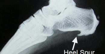
An examination of the foot is enough for the health care provider to diagnose flat foot. However, the cause must be determined. If an arch develops when the patient stands on his or her toes, the flat foot is called flexible and no treatment or further work-up is necessary. If there is pain associated with the foot or if the arch does not develop with toe-standing, x-rays are necessary. If a tarsal coalition is suspected, a CT scan is often ordered. If a posterior tibial tendon injury is suspected, your health care provider may recommend an MRI.

Can you fix a fallen arch?

Non Surgical Treatment

If you have fallen arches, but you are not experiencing any symptoms, then you probably do not need to seek treatment. If you are experiencing discomfort due to fallen arches, there are several treatment options. These treatment options include elevating the feet and applying ice to ease discomfort and reduce swelling, rest, exercises to stretch the feet, physical therapy, medication, such as anti-inflammatories, steroid injections and orthotic devices or customised arch supportsto wear in the shoes. If you have fallen arches and periodically experience pain related to that condition, it is a good idea to get orthotic devicesor custom arch supports, to wear in your shoes. The other treatment options, like medication and ice, will help to ease pain from fallen arches after you have already begun to experience pain. However, orthotic devices or(custom arch supports)can help to prevent pain from occurring at all. This preventative measure helps many people with fallen arches to avoid pain and prevent worsening of their condition. In severe cases of fallen arches, surgery may be required to correct the problem. You can also help to prevent pain and exacerbation of fallen arches by reducing your risk factors. If you are overweight, try to lose weight. Even a small weight loss can reduce the pressure on your feet significantly. If you are diabetic, manage your blood sugar as best as possible. Losing weight often also improves the condition of diabetics. You should also avoid high-impact activities, like running on the road, tennis, and sports that involve jumping. Try a gentler form of exercise, like swimming, instead. If you have fallen arches, orthotic devices or(custom arch supports)are an important component of your treatment and can help to prevent pain.

Surgical Treatment

Adult Acquired Flat Feet

Surgical correction is dependent on the severity of symptoms and the stage of deformity. The goals of surgery are to create a more functional and stable foot. There are multiple procedures available to the surgeon and it may take several to correct a flatfoot deformity. Stage one deformities usually respond to conservative or non-surgical therapy such as anti-inflammatory medication, casting, functional orthotics or a foot ankle orthosis called a Richie Brace. If these modalities are unsuccessful surgery is warranted. Usually surgical treatment begins with removal of inflammatory tissue and repair of the posterior tibial tendon. A tendon transfer is performed if the posterior tibial muscle is weak or the tendon is badly damaged. The most commonly used tendon is the flexor digitorum longus tendon. This tendon flexes or moves the lesser toes downward. The flexor digitorum longus tendon is utilized due to its close proximity to the posterior tibial tendon and because there are minimal side effects with its loss. The remainder of the tendon is sutured to the flexor hallucis longus tendon that flexes the big toe so that little function is loss. Stage two deformities are less responsive to conservative therapies that can be effective in mild deformities. Bone procedures are necessary at this stage in order to recreate the arch and stabilize the foot. These procedures include isolated fusion procedures, bone grafts, and/or the repositioning of bones through cuts called osteotomies. The realigned bones are generally held in place with screws, pins, plates, or staples while the bone heals. A tendon transfer may or may not be utilized depending on the condition of the posterior tibial tendon. Stage three deformities are better treated with surgical correction, in healthy patients. Patients that are unable to tolerate surgery or the prolonged healing period are better served with either arch supports known as orthotics or bracing such as the Richie Brace. Surgical correction at this stage usually requires fusion procedures such as a triple or double arthrodesis. This involves fusing the two or three major bones in the back of the foot together with screws or pins. The most common joints fused together are the subtalar joint, talonavicular joint, and the calcaneocuboid joint. By fusing the bones together the surgeon is able to correct structural deformity and alleviate arthritic pain. Tendon transfer procedures are usually not beneficial at this stage. Stage four deformities are treated similarly but with the addition of fusing the ankle joint.

How To Assess For Leg Length Discrepancy

7/2/2017

 
Overview

Some people have an ?apparent? LLD which may make the affected leg seem longer than the other leg. There are several factors that can contribute to this feeling. Most commonly, contractures or shortening of the muscles surrounding the hip joint and pelvis make the involved leg feel longer, even when both legs are really the same length. Additionally, contractures of the muscles around the lower back from spinal disorders (i.e. arthritis, spinal stenosis), curvatures of the spine from scoliosis, and deformities of the knee or ankle joint can make one leg seem longer or shorter. In the general public, some people have an ?apparent LLD? as long as one half inch but usually don?t notice it because the LLD occurs over time. A ?true? LLD is where one leg is actually longer than the other. Patients can have unequal leg lengths of 1/4? to 1/2? and never feel it too! You can also have combinations of ?True? and ?Apparent? LLDs. During total hip replacement surgery, the surgeon may ?lengthen? the involved leg by stretching the muscles and ligaments that were contracted, as well as by restoring the joint space that had become narrowed from the arthritis. This is usually a necessary part of the surgery because it also provides stability to the new hip joint. Your surgeon takes measurements of your leg lengths on x-ray prior to surgery. Your surgeon always aims for equal leg lengths if at all possible and measures the length of your legs before and during surgery in order to achieve this goal. Occasionally, surgeons may need to lengthen the operable leg to help improve stability and prevent dislocations as well improve the muscle function around the hip.Leg Length Discrepancy

Causes

There are many causes of leg length discrepancy. Structural inequality is due to interference of normal bone growth of the lower extremity, which can occur from trauma or infection in a child. Functional inequality has many causes, including Poliomyelitis or other paralytic deformities can retard bone growth in children. Contracture of the Iliotibial band. Scoliosis or curvature of the spine. Fixed pelvic obliquity. Abduction or flexion contraction of the hip. Flexion contractures or other deformities of the knee. Foot deformities.

Symptoms

The symptoms of limb deformity can range from a mild difference in the appearance of a leg or arm to major loss of function of the use of an extremity. For instance, you may notice that your child has a significant limp. If there is deformity in the extremity, the patient may develop arthritis as he or she gets older, especially if the lower extremities are involved. Patients often present due to the appearance of the extremity (it looks different from the other side).

Diagnosis

The doctor carefully examines the child. He or she checks to be sure the legs are actually different lengths. This is because problems with the hip (such as a loose joint) or back (scoliosis) can make the child appear to have one shorter leg, even though the legs are the same length. An X-ray of the child?s legs is taken. During the X-ray, a long ruler is put in the image so an accurate measurement of each leg bone can be taken. If an underlying cause of the discrepancy is suspected, tests are done to rule it out.

Non Surgical Treatment

Treatments for limb-length discrepancies and differences vary, depending on the cause and severity of the condition. At Gillette, our orthopedic surgeons are experts in typical and atypical growth and development. Our expertise lets us plan treatments that offer a lifetime of benefits. Treatments might include monitoring growth and development, providing noninvasive treatments or therapy, and providing a combination of orthopedic surgical procedures. To date, alternative treatments (such as chiropractic care or physical therapy) have not measurably altered the progression of or improved limb-length conditions. However, children often have physical or occupational therapy to address related conditions, such as muscle weakness or inflexibility, or to speed recovery following a surgical procedure. In cases where surgical treatment isn?t necessary, our orthopedists may monitor patients and plan noninvasive treatments, such as, occupational therapy, orthoses (braces) and shoe inserts, physical therapy, prostheses (artificial limbs).

LLL Shoe Insoles

leg length discrepancy lifts

Surgical Treatment

The type of surgery depends on the type of problem. Outpatient procedures may be used to alter the growth of the limb. This is often done through small incisions. If an outpatient procedure is done, your child can continue with most regular activities. Other times, surgery may be very involved and require the use of an external device that is attached to the limb with pins and wires. This device may be left on for months to correct the deformity or lengthen the leg. If this type of surgery is required, your child will be making weekly visits to Cincinnati Children's.

Everything You Want To Understand About Heel Soreness

6/29/2017

 
Overview

Painful Heel

Heel pain is usually felt either under the heel or just behind it. Heel pain has a prevalence of 3.6%. US studies estimate that 7% of older adults report tenderness under the heel. Plantar fasciitis is estimated to account for 8% of all running-related injuries. There are 26 bones in the human foot, of which the heel is the largest. Pain typically comes on gradually, with no injury to the affected area. It is often triggered by wearing a flat shoe. In most cases the pain is under the foot, towards the front of the heel. The majority of patients recover with conservative treatments within months. Home care such as rest, ice, proper-fitting footwear and foot supports are often enough to ease heel pain. To prevent heel pain, it's recommended to reduce the stress on that part of the body.

Causes

Long standing inflammation causes the deposition of calcium at the point where the plantar fascia inserts into the heel. This results in the appearance of a sharp thorn like heel spur on x-ray. The heel spur is asymptomatic (not painful), the pain arises from the inflammation of the plantar fascia.

Symptoms

Symptoms of plantar fasciitis vary, but the classic symptom is pain after rest--when you first get out of bed in the morning, or when you get up after sitting down for a while during the day. The pain usually diminishes after a few minutes of walking, sometimes even disappearing, but the pain is commonly felt again the longer you're on the foot. Fasciitis can be aggravated by shoes that lack appropriate support, especially in the arch area, and by the chronic irritation of long-periods of standing, especially on concrete, by being overweight. It doesn't help that fascia doesn't heal particularly quickly because it has relatively poor circulation (which is why it's white in colour).

Diagnosis

To arrive at a diagnosis, the foot and ankle surgeon will obtain your medical history and examine your foot. Throughout this process the surgeon rules out all the possible causes for your heel pain other than plantar fasciitis. In addition, diagnostic imaging studies such as x-rays or other imaging modalities may be used to distinguish the different types of heel pain. Sometimes heel spurs are found in patients with plantar fasciitis, but these are rarely a source of pain. When they are present, the condition may be diagnosed as plantar fasciitis/heel spur syndrome.

Non Surgical Treatment

Curing posterior heel pain requires calming the inflammation, resting the foot and increasing calf flexibility. Ice therapy and anti-inflammatory medications can be used to reduce the inflammation. Stopping exercises that stress the calf and Achilles is important. This includes walking, running and the use of stair climbers and elliptical machines. Placing a heel lift in each shoe can reduce some of the excess strain on the back of the heel. Stretching exercises to increase calf flexibility are important in curing this problem and preventing its recurrence. Wearing running shoes also provides good foot support and helps with this condition. Sometimes a walking boot is used to immobilize the ankle and let the area completely rest. Physical therapy is sometimes ordered to help reduce the inflammation and pain and to help improve the flexibility of the calf muscles. Occasionally these measures fail to relieve the pain and surgery may be needed. The surgical procedure involves removing bone spurs and repairing any damage to the tendon.

Surgical Treatment

With the advancements in technology and treatments, if you do need to have surgery for the heel, it is very minimal incision that?s done. And the nice thing is your recovery period is short and you should be able to bear weight right after the surgery. This means you can get back to your weekly routine in just a few weeks. Recovery is a lot different than it used to be and a lot of it is because of doing a minimal incision and decreasing trauma to soft tissues, as well as even the bone. So if you need surgery, then your recovery period is pretty quick.

Why do I have pain in my heel?

Prevention

Heel Pain

Being overweight can place excess pressure and strain on your feet, particularly on your heels. Losing weight, and maintaining a healthy weight by combining regular exercise with a healthy, balanced diet, can be beneficial for your feet. Wearing appropriate footwear is also important. Ideally, you should wear shoes with a low to moderate heel that supports and cushions your arches and heels. Avoid wearing shoes with no heels.

What Causes Mortons Neuroma

6/5/2017

 
Overview

Morton neuromaMorton's neuroma is an enlarged nerve that usually occurs in the third interspace, which is between the third and fourth toes. To understand Morton's neuroma further, it may be helpful to look at the anatomy of the foot. Problems often develop in the third interspace because part of the lateral plantar nerve combines with part of the medial plantar nerve here. When the two nerves combine, they are typically larger in diameter than those going to the other toes. Also, the nerve lies in subcutaneous tissue, just above the fat pad of the foot, close to an artery and vein. Above the nerve is a structure called the deep transverse metatarsal ligament. This ligament is very strong, holds the metatarsal bones together, and creates the ceiling of the nerve compartment. With each step, the ground pushes up on the enlarged nerve and the deep transverse metatarsal ligament pushes down. This causes compression in a confined space.

Causes

In many cases, a neuroma may develop as a result of excessive loading on the front of the foot. Sometimes, a patient?s anatomic alignment in the forefoot contributes to the overload. There may be some cases where the neuroma develops spontaneously, for no obvious reason. However, once the nerve is irritated, pressure from walking, and from the adjacent bony prominences (metatarsal heads), as well as from the intermetatarsal ligament that binds the heads together, all may contribute to persistent pain. Repetitive pressure on the nerve causes localized injury with resulting scarring and fibrosis of the nerve. This leads to symptoms in the distribution of the nerve.

Symptoms

Neuroma pain is classically described as a burning pain in the forefoot. It can also be felt as an aching or shooting pain in the forefoot. Patients with this problem frequently say they feel like they want to take off their shoes and rub their foot. This pain may occur in the middle of a run or at the end of a long run. If your shoes are quite tight or the neuroma is very large, the pain may be present even when walking. Occasionally a sensation of numbness is felt in addition to the pain or even before the pain appears.

Diagnosis

Plain x-rays of the foot may demonstrate that one or more of the metatarsals are long (Figure #5). Not uncommonly, the second and/or third metatarsal may be long relative to the third or fourth. This can create a situation where excessive load is occurring in and around the vicinity of the interdigital nerve.

Non Surgical Treatment

Wear shoes with plenty of room for the toes to move, low heels, and laces or buckles that allow for width adjustment. Wear shoes with thick, shock-absorbent soles, as well as proper insoles that are designed to keep excessive pressure off of the foot. High-heeled shoes over two inches tall should be avoided whenever possible because they place undue strain on the forefoot. Resting the foot and massaging the affected area can temporarily alleviate neuroma pain. Use an ice pack to help to dull the pain and improve comfort. Use over-the-counter shoe pads. These pads can relieve pressure around the affected area.Morton neuroma

Surgical Treatment

Majority of publications including peer review journal articles, surgical technique description and textbooks promote surgical excision as a gold standard treatment. Surgical excision is described as the most definitive mode of treatment for symptomatic Morton?s neuroma with reported success rates varying between 79% and 93%. Various surgical techniques are described, essentially categorised as dorsal versus plantar incision approaches. Beyond this the commonest technical variation described as influencing the outcome of surgery involves burying and anchoring transacted nerve into soft tissue such as muscle.

Leg Length Discrepancy And Shoe Lifts

2/23/2016

 
There are two unique variations of leg length discrepancies, congenital and acquired. Congenital means you are born with it. One leg is structurally shorter in comparison to the other. Through developmental periods of aging, the brain senses the walking pattern and recognizes some variance. Your body usually adapts by tilting one shoulder to the "short" side. A difference of under a quarter inch isn't blatantly excessive, does not need Shoe Lifts to compensate and typically does not have a profound effect over a lifetime.

 <a href="http://shockingdeity5346.exteen.com/20150429/leg-length-discrepancy-treatment-after-hip-replacement">Shoe Lifts</a>

Leg length inequality goes largely undiagnosed on a daily basis, however this condition is easily solved, and can eradicate many instances of chronic back pain.

Treatment for leg length inequality commonly involves Shoe Lifts. They are cost-effective, typically priced at under twenty dollars, in comparison to a custom orthotic of $200 and up. Differences over a quarter inch can take their toll on the spine and should probably be compensated for with a heel lift. In some cases, the shortage can be so extreme that it requires a full lift to both the heel and sole of the shoe.

Chronic back pain is easily the most widespread health problem impacting people today. Around 80 million men and women have problems with back pain at some stage in their life. It's a problem which costs companies vast amounts of money year after year on account of time lost and productivity. Fresh and more effective treatment methods are always sought after in the hope of decreasing the economic influence this condition causes.

Leg Length Discrepancy Shoe Lift

Men and women from all corners of the earth suffer the pain of foot ache as a result of leg length discrepancy. In most of these situations Shoe Lifts might be of worthwhile. The lifts are capable of easing any discomfort in the feet. Shoe Lifts are recommended by numerous professional orthopaedic physicians.

To be able to support the human body in a balanced manner, the feet have a critical task to play. Irrespective of that, it can be the most neglected region in the human body. Many people have flat-feet meaning there may be unequal force exerted on the feet. This causes other areas of the body including knees, ankles and backs to be impacted too. Shoe Lifts make sure that correct posture and balance are restored.

Just What Is Calcaneal Spur

9/28/2015

 
Posterior Calcaneal Spur

Overview

Heel spurs are abnormal bony growths that develop at the back of or under the heel. Inflammation around a spur, more so than the spur itself, can cause significant pain. Fortunately, symptoms can be eased with non-surgical treatments for the vast majority of people.

Causes

A heel spur usually develops as a result of wear and tear over time, which leads to the degeneration of connective tissue called fascia. Standing for prolonged periods and wearing shoes that do not provide the right type of arch support can also lead to connective tissue damage in the heel. The body attempts to repair the damaged tissue by delivering calcium to the affected region, but sometimes too much calcium begins to accumulate and this results in painful plantar fasciitis.

Posterior Calcaneal Spur

Symptoms

Bone spurs may cause sudden, severe pain when putting weight on the affected foot. Individuals may try to walk on their toes or ball of the foot to avoid painful pressure on the heel spur. This compensation during walking or running can cause additional problems in the ankle, knee, hip, or back.

Diagnosis

A Diagnosis of Heel Spur Syndrome is a very common reason for having heel pain. Heel pain may be due to other types of conditions such as tendonitis, Haglund's Deformity, Stress Fracture, Tarsal Tunnel Syndrome, or low back problems. A more common condition in children is Sever's Disease. The diagnosis is usually made with a combination of x-ray examination and symptoms.

Non Surgical Treatment

By reducing excessive motion and controlling and supporting the foot during physical activities an orthotic can help to limit how far the plantar fascia is pulled or torn away from the heel. A Heel Spur pad can be offered- which is a pad designed to take pressure off the spur. If the problem persists, consult your foot doctor.

Surgical Treatment

Usually, heel spurs are curable with conservative treatment. If not, heel spurs are curable with surgery, although there is the possibility of them growing back. About 10% of those who continue to see a physician for plantar fascitis have it for more than a year. If there is limited success after approximately one year of conservative treatment, patients are often advised to have surgery.

Prevention

In order to prevent heel spurs, it?s important that you pay attention to the physical activities you engage in. Running or jogging on hard surfaces, such as cement or blacktop, is typical for competitive runners, but doing this for too long without breaks can lead to heel spurs and foot pain. Likewise, the shoes you wear can make a big difference in whether or not you develop heel spurs. Have your shoes and feet checked regularly by our Dallas podiatrist to ensure that you are wearing the proper equipment for the activities. Regular checkups with a foot and ankle specialist can help avoid the development of heel spurs.

What Is The Most Beneficial Strategy For Calcaneal Spur

9/26/2015

 
Calcaneal Spur

Overview

A heel spur is a calcium deposit on the underside of the heel bone, often caused by strain on foot muscles and ligaments. Heel spurs are common among athletes but also tend to develop as we age, as flexibility decreases. Heel spurs can be painful when associated with plantar fasciitis, an inflammation of the connective tissue that runs along the bottom of the foot and connects the heel bone to the ball of the foot.

If left untreated, the mild aches associated with this condition can evolve into chronic pain. And as you try to compensate for the pain, your gait may change, which could impact your knee, hip and back.

Causes

Heel spurs are bony outgrowths positioned where the plantar fascia tissue attaches to the heel bone (the calcaneus). Heel spurs seldom cause pain. It is the inflamed tissue surrounding the spur that causes the pain. The Latin meaning of Plantar Fasciitis is, ?Inflammation of Plantar Fascia.? The plantar fascia is a long, thick and very tough band of tissue beneath your foot that provides arch support. It also connects your toes to your heel bone. Each time you take a step, the arch slightly flattens to absorb impact. This band of tissue is normally quite strong and flexible but unfortunately, circumstances such as undue stress, being overweight, getting older or having irregularities in your foot dynamics can lead to unnatural stretching and micro-tearing of the plantar fascia. This causes pain and swelling at the location where the plantar fascia attaches to the heel bone. As the fascia continually pulls at the heel bone, the constant irritation eventually creates a bony growth on the heel. This is called a heel spur.

Inferior Calcaneal Spur

Symptoms

An individual with the lower legs turning inward, a condition called genu valgus or "knock knees," can have a tendency toward excessive pronation. This can lead to a fallen arch and problems with the plantar fascia and heel spurs. Women tend to suffer from this condition more than men. Heel spurs can also result from an abnormally high arch. Other factors leading to heel spurs include a sudden increase in daily activities, an increase in weight, or a thinner cushion on the bottom of the heel due to old age. A significant increase in training intensity or duration may cause inflammation of the plantar fascia. High-heeled shoes, improperly fitted shoes, and shoes that are too flexible in the middle of the arch or bend before the toe joints will cause problems with the plantar fascia and possibly lead to heel spurs.

Diagnosis

A heel spur is often seen on X-ray as a bony protrusion, which can vary in size. However, because a Heel Spur only indicates increased load on the plantar fascia, and not pain, an ultra sound may be required to assess other actual cause of the heel pain such and may include checking to see if the plantar fascia is inflamed or degenerated.

Non Surgical Treatment

By reducing excessive motion and controlling and supporting the foot during physical activities an orthotic can help to limit how far the plantar fascia is pulled or torn away from the heel. A Heel Spur pad can be offered- which is a pad designed to take pressure off the spur. If the problem persists, consult your foot doctor.

Surgical Treatment

When chronic heel pain fails to respond to conservative treatment, surgical treatment may be necessary. Heel surgery can provide relief of pain and restore mobility. The type of procedure used is based on examination and usually consists of releasing the excessive tightness of the plantar fascia, called a plantar fascia release. Depending on the presence of excess bony build up, the procedure may or may not include removal of heel spurs. Similar to other surgical interventions, there are various modifications and surgical enhancements regarding surgery of the heel.

Bursa Foot Indications

8/23/2015

 
Overview

Bursitis is the painful inflammation of the bursa, a padlike sac found in areas subject to friction. Bursae cushion the movement between the bones, tendons and muscles near the joints. Bursitis is most often caused by repetitive movement and is known by several common names including weaver's bottom, clergyman's knee, and miner's elbow, depending on the affected individual's occupation and area of injury.

Causes

Retrocalcaneal bursitis is generally caused by local trauma from poorly designed shoes. Patients complain of posterolateral heel pain and may have a posterior heel prominence (?pump bump?), as well as local swelling and tenderness over the Achilles tendon. Pain is increased by squeezing the bursa from side to side and anterior to the Achilles. A heel lift and open-back shoes help alleviate pressure.

Symptoms

Pain and tenderness usually develop slowly over time. Applying pressure to the back of the heel can cause pain. Wearing shoes may become uncomfortable. The back of the heel may feel achy. Pain is exacerbated when the foot is pointed or flexed, because the swollen bursa can get squeezed. A person with retrocalcaneal bursitis may feel pain when standing on their toes. Fever or chills in addition to other bursitis symptoms can be a sign of septic bursitis. Though uncommon, septic retrocalcaneal bursitis is a serious condition, and patients should seek medical care to ensure the infection does not spread.

Diagnosis

On physical examination, patients have tenderness at the site of the inflamed bursa. If the bursa is superficial, physical examination findings are significant for localized tenderness, warmth, edema, and erythema of the skin. Reduced active range of motion with preserved passive range of motion is suggestive of bursitis, but the differential diagnosis includes tendinitis and muscle injury. A decrease in both active and passive range of motion is more suggestive of other musculoskeletal disorders. In patients with chronic bursitis, the affected limb may show disuse atrophy and weakness. Tendons may also be weakened and tender.

Non Surgical Treatment

Cold compresses can help reduce the initial swelling and pain in acute (short-term but severe) soft tissue conditions. Cold therapy is usually most effective during the first 48 hours after swelling begins. Guidelines for cold therapy include. Use a cold gel pack, a bag filled with ice cubes, or even a bag of frozen vegetables. Wrap the pack in a towel if the cold temperature is too painful. Place the cold pack over the area for 20 minutes, three to four times a day. Rub an ice cube over smaller painful areas for a short time. After 48 hours, or for chronic (long-term) pain, dry or moist heat may be more helpful than cold compresses. Follow these guidelines. Use a hot pack, a heating pad, or a damp towel heated in the microwave (make sure it's not too hot or it may burn your skin). Place a hot pack over the painful area for 15-20 minutes, three to four times a day. Never use analgesic creams or rubs with heat packs because the combination could severely burn your skin. Take a warm shower or bath.

Surgical Treatment

Surgery. Though rare, particularly challenging cases of retrocalcaneal bursitis might warrant a bursectomy, in which the troublesome bursa is removed from the back of the ankle. Surgery can be effective, but operating on this boney area can cause complications, such as trouble with skin healing at the incision site. In addition to removing the bursa, a doctor may use the surgery to treat another condition associated with the retrocalcaneal bursitis. For example, a surgeon may remove a sliver of bone from the back of the heel to alter foot mechanics and reduce future friction. Any bone spurs located where the Achilles attaches to the heel may also be removed. Regardless of the conservative treatment that is provided, it is important to wait until all pain and swelling around the back of the heel is gone before resuming activities. This may take several weeks. Once symptoms are gone, a patient may make a gradual return to his or her activity level before their bursitis symptoms began. Returning to activities that cause friction or stress on the bursa before it is healed will likely cause bursitis symptoms to flare up again.

Prevention

Maintain proper form when exercising, good flexibility, and strength around the ankle to help prevent this condition from arising. Proper stretching of the achilles tendon helps prevent injury.

New Hammertoe Surgery

6/23/2015

 
HammertoeOverview

A hammertoes is a deformity of the second, third or fourth toes. In this condition, the toe is bent at the middle joint, so that it resembles a hammer. Initially, hammer toes are flexible and can be corrected with simple measures but, if left untreated, they can become fixed and require surgery. People with hammer toe may have corns or calluses on the top of the middle joint of the toe or on the tip of the toe. They may also feel pain in their toes or feet and have difficulty finding comfortable shoes.

Causes

Hammer toe usually affects the second toe. However, it may also affect the other toes. The toe moves into a claw-like position. The most common cause of hammer toe is wearing short, narrow shoes that are too tight. The toe is forced into a bent position. Muscles and tendons in the toe tighten and become shorter. Hammer toe is more likely to occur in women who wear shoes that do not fit well or have high heels and children who keep wearing shoes they have outgrown. The condition may be present at birth (congenital) or develop over time. In rare cases, all of the toes are affected. This may be caused by a problem with the nerves or spinal cord.

HammertoeSymptoms

Common symptoms of hammertoes include pain or irritation of the affected toe when wearing shoes. corns and calluses (a buildup of skin) on the toe, between two toes, or on the ball of the foot. Corns are caused by constant friction against the shoe. They may be soft or hard, depending upon their location. Inflammation, redness, or a burning sensation. Contracture of the toe. In more severe cases of hammertoe, open sores may form.

Diagnosis

The exam may reveal a toe in which the near bone of the toe (proximal phalanx) is angled upward and the middle bone of the toe points in the opposite direction (plantar flexed). Toes may appear crooked or rotated. The involved joint may be painful when moved, or stiff. There may be areas of thickened skin (corns or calluses) on top of or between the toes, a callus may also be observed at the tip of the affected toe beneath the toenail. An attempt to passively correct the deformity will help elucidate the best treatment option as the examiner determines whether the toe is still flexible or not. It is advisable to assess palpable pulses, since their presence is associated with a good prognosis for healing after surgery. X-rays will demonstrate the contractures of the involved joints, as well as possible arthritic changes and bone enlargements (exostoses, spurs). X-rays of the involved foot are usually performed in a weight-bearing position.

Non Surgical Treatment

Apply a commercial, nonmedicated hammertoe pad around the bony prominence of the hammertoe. This will decrease pressure on the area. Wear a shoe with a deep toe box. If the hammertoe becomes inflamed and Hammer toe painful, apply ice packs several times a day to reduce swelling. Avoid heels more than two inches tall. A loose-fitting pair of shoes can also help protect the foot while reducing pressure on the affected toe, making walking a little easier until a visit to your podiatrist can be arranged. It is important to remember that, while this treatment will make the hammertoe feel better, it does not cure the condition. A trip to the podiatric physician?s office will be necessary to repair the toe to allow for normal foot function. Avoid wearing shoes that are too tight or narrow. Children should have their shoes properly fitted on a regular basis, as their feet can often outgrow their shoes rapidly. See your podiatric physician if pain persists.

Surgical Treatment

Sometimes when the joints are removed the two bones become one as they are fused in a straightened position. Many times one toe will be longer than another and a piece of bone is removed to bring the toes in a more normal length in relation to each other. Sometimes tendons will be lengthened, or soft tissue around the joints will be cut or rebalanced to fix the deformity. Angular corrections may also be needed. The surgeon may place fixation in your foot as it heals which may include a pin, or wires.

Over-Pronation

5/31/2015

 
Overview

Over-pronation, or flat feet, is a common biomechanical problem that occurs in the walking process when a person?s arch collapses upon weight bearing. This motion can cause extreme stress or inflammation on the plantar fascia, possibly causing severe discomfort and leading to other foot problems.Foot Pronation

Causes

Over-pronation may happen because the tissue that attaches to your foot bones is loose. You may be born with this problem or it may result from injuries or overuse, like from too much running.

Symptoms

Overpronation causes alterations in proper muscle recruitment patterns leading to tightness in the outside of the ankle (lateral gastrocnemius, soleus, and peroneals). This tightness can lead to weakness in the opposing muscles such as the medial gastrocnemius, anterior tibialis, and posterior tibialis. If these muscles are weak, they will not be able to keep the knee in proper alignment, causing the valgus position. All this tightness and weakness can cause pain within the ankle, calf, and knee region. And it can send imbalance and pain all the way up to the upper back, if deep core strength is lacking and can't hold the pelvis in neutral.

Diagnosis

Firstly, look at your feet in standing, have you got a clear arch on the inside of the foot? If there is not an arch and the innermost part of the sole touches the floor, then your feet are over-pronated. Secondly, look at your running shoes. If they are worn on the inside of the sole in particular, then pronation may be a problem for you. Thirdly, try the wet foot test. Wet your feet and walk along a section of paving and look at the footprints you leave. A normal foot will leave a print of the heel, connected to the forefoot by a strip approximately half the width of the foot on the outside of the sole. If you?re feet are pronated there may be little distinction between the rear and forefoot, shown opposite. The best way to determine if you over pronate is to visit a podiatrist or similar who can do a full gait analysis on a treadmill or using forceplates measuring exactly the forces and angles of the foot whilst running. It is not only the amount of over pronation which is important but the timing of it during the gait cycle as well that needs to be assessed.Over-Pronation

Non Surgical Treatment

Studies have shown that the most effective way to dexrease a high Q angle and lower the biomechanical stresses on the knee joint is to prevent excessive pronation with custom-maflexible orthotics. One study found that using soft corrective orthotics was more effective in reduknee pain than a traditional exercise program. A more recent study showed that Q angle asymmetries, secondary to excessive pronation affecting knee alignment, can be effectivecontrolled or corrected utilizing custom-made, flexible orthotics. Another project involving meof a running club determined that 75% of those using orthotics eliminated or greatly reduced pain in the feet, ankles, shins, knees and hips

Prevention

Firstly, a thorough and correct warm up will help to prepare the muscles and tendons for any activity or sport. Without a proper warm up the muscles and tendons around your feet, ankles and lower legs will be tight and stiff. There will be limited blood flow to the lower legs, which will result in a lack of oxygen and nutrients for those muscles. Click here for a detailed explanation of how, why and when to perform your warm up. Secondly, flexible muscles are extremely important in the prevention of most ankle and lower leg injuries. When muscles and tendons are flexible and supple, they are able to move and perform without being over stretched. If however, your muscles and tendons are tight and stiff, it is quite easy for those muscles and tendons to be pushed beyond their natural range of motion. To keep your muscles and tendons flexible and supple, it is important to undertake a structured stretching routine.
<<Previous

    Author

    Write something about yourself. No need to be fancy, just an overview.

    Archives

    July 2017
    June 2017
    February 2016
    September 2015
    August 2015
    June 2015
    May 2015
    March 2015
    February 2015
    June 2014

    Categories

    All
    Foot Conditions

    RSS Feed

Powered by Create your own unique website with customizable templates.